


Product Areas
-

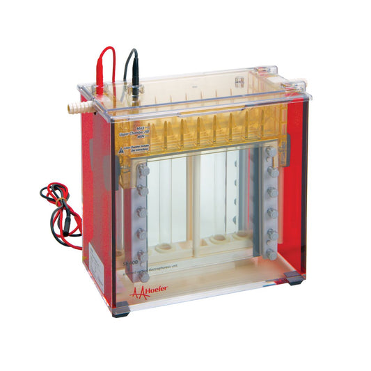
Protein Electrophoresis
Our globally-celebrated protein electrophoresis products, including our SE series of Mini and...
-

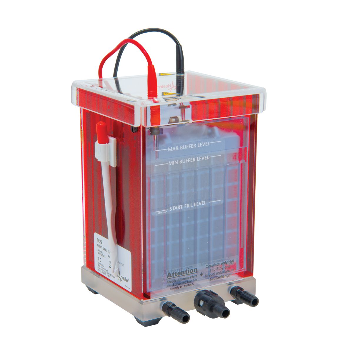
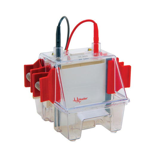
Nucleic Acid Electrophoresis
Hoefer's celebrated line of Nucleic Acid Electrophoresis products, including our popular SUB...
-

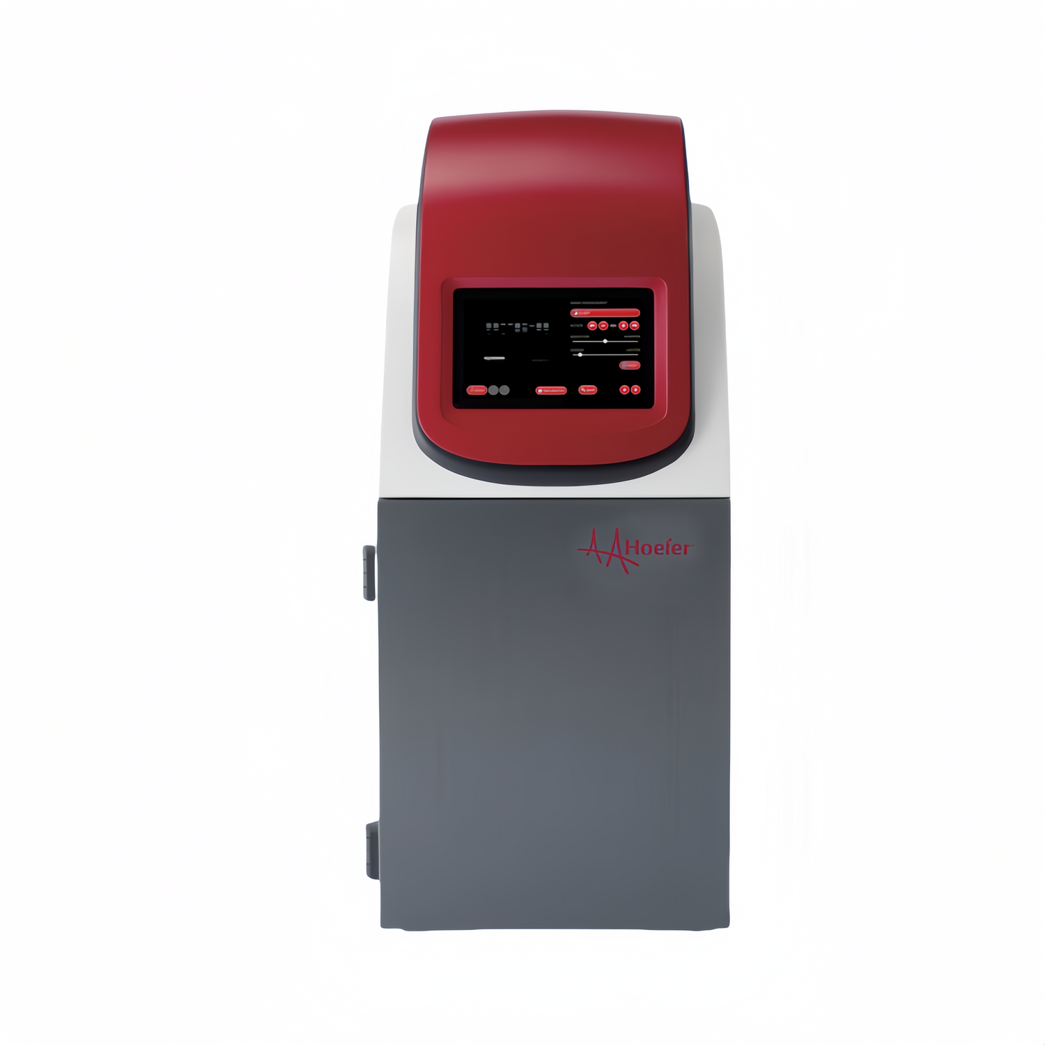
Power Supply
Economical, reliable power supply units for Hoefer's gel electrophoresis, blotting, and other...
-

Sample Storage Solutions
All of Hoefer's latest products in our groundbreaking end-to-end solution for complete,...